

Please fill in the form below !




Revision rhinoplasty for a visible nasal implant is intended to address implant show-through after a previous high-bridge rhinoplasty, where the implant contour becomes noticeable beneath the skin and may be accompanied by redness, shiny or tight skin, or discomfort.
This guide from EMCAS Aesthetic Hospital explains why implant visibility can occur after high-bridge rhinoplasty, the common contributing factors, how revision surgery is performed, and the expected cost.
In recent years, a tall, straight nasal bridge with a refined tip has become a popular aesthetic preference, particularly among younger patients who prefer a more defined facial profile.
However, not every nose can safely accommodate excessive bridge height. When an implant creates more projection than the skin and soft-tissue envelope can adequately cover, the risk of implant visibility, redness, and progressive skin thinning may increase over time.
For this reason, some patients who previously chose a very high nasal bridge later seek revision rhinoplasty to improve implant coverage, facial harmony, and long-term structural stability.
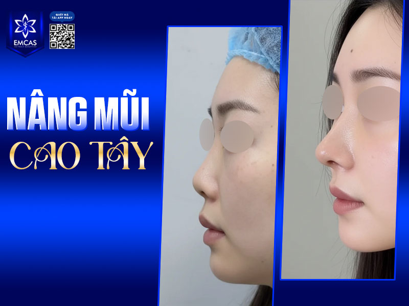
A visible nasal implant occurs when the outline or edge of an implant placed along the nasal bridge becomes noticeable beneath the skin, creating a hard or unnatural contour. This can occur when the implant is too prominent for the patient’s original nasal anatomy and soft-tissue coverage.
At first, the nose may appear tall and straight with a sharply defined profile. Over time, however, continued tension on thin skin and inadequate soft-tissue coverage can make the implant contour more visible, especially from the side or under strong lighting. In more advanced cases, problems may include capsular contracture, implant migration, bridge deviation, or a risk of skin breakdown at the nasal tip.
Nasal implant visibility can be influenced by several factors, including implant height and material, surgical technique, individual soft-tissue characteristics, and postoperative care. Identifying the likely cause is an important part of planning revision surgery and reducing the risk of recurrence.
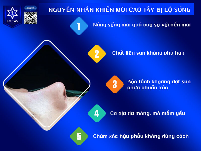
Revision rhinoplasty for a visible nasal implant is generally more complex than primary rhinoplasty because the nose has already been operated on. The previous implant remains in place, while the soft tissue may be thinner, scarred, adherent, or weakened. At EMCAS, the revision process includes the following steps:
Step 1: Consultation and nasal assessment
The surgeon evaluates the degree of implant visibility, skin thickness, soft-tissue scarring or adhesions, and the condition, position, and material of the previous implant. This assessment helps determine whether the implant needs to be completely removed or whether partial correction may be appropriate.
The surgeon also discusses a more individualized nasal shape based on the patient’s existing anatomy, with the aim of avoiding excessive bridge height that could place renewed tension on the soft tissue.
Step 2: Anesthesia
The revision procedure is performed in a sterile operating room. General anesthesia is used so the patient does not experience pain or discomfort during surgery.
Step 3: Removal of the previous implant and treatment of the implant pocket
In many cases, the previous synthetic implant is removed completely. If there is a thick or contracted capsule, the surgeon may release or treat the scar tissue to reduce continued compression.
The implant pocket is then cleaned and recreated at an appropriate anatomical plane. Careful pocket preparation is important because an inadequately treated pocket may contribute to recurrent problems or bridge deviation.
Step 4: Structural revision and placement of a new implant or graft
A replacement material is selected with attention to appropriate height and flexibility. The surgeon creates the pocket and positions the material at the intended anatomical plane to improve soft-tissue coverage and reduce the likelihood of recurrent implant visibility.
Autologous cartilage, such as ear cartilage or septal cartilage, may also be used to support or cover the nasal tip and improve tissue coverage. The objective is not only to reduce implant show-through but also to create a stable, softer, and more natural-looking nasal structure.
Step 5: Postoperative monitoring and care
After surgery, the patient is monitored in the recovery area and stays at the hospital for at least one day before going home. Following postoperative instructions and attending scheduled follow-up visits are important for monitoring healing and identifying any concerning signs early.
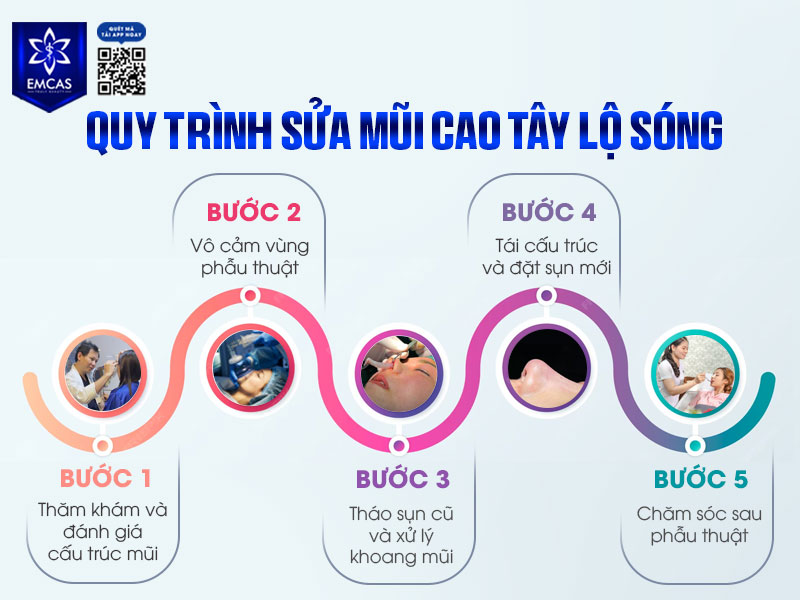
At EMCAS Aesthetic Hospital, revision rhinoplasty for a visible nasal implant starts from VND 23 million. The final cost varies by case and depends on the condition of the nose and the treatment required. For an individualized quotation, contact EMCAS at 0903 082 836 or 0979 223 636.
Revision rhinoplasty for implant visibility can be more complex than primary rhinoplasty and requires careful assessment of the nasal anatomy, previous implant, scar tissue, and soft-tissue coverage. At EMCAS Aesthetic Hospital, the procedure is planned on an individual basis with two main goals: addressing the visible implant contour and rebuilding a nasal shape that is proportionate to the patient’s facial anatomy.
The EMCAS medical team has experience in primary and revision rhinoplasty, including cases involving implant visibility, capsular contracture, and thin soft-tissue coverage. Detailed anatomical assessment and surgical planning are important parts of managing these more complex cases.
Each revision case at EMCAS begins with an examination of the current nasal structure. The surgeon evaluates skin thickness, the degree of implant visibility, the condition of the previous synthetic implant, capsular contracture, and any soft-tissue damage.
Based on these findings, an individualized revision plan is developed rather than applying the same approach to every patient.
The procedure is performed in a sterile operating room with anesthesia and postoperative monitoring. This is especially important in revision cases because surgery may take longer and the nasal tissues have already undergone previous surgical intervention.
EMCAS Aesthetic Hospital has diagnostic and imaging equipment including a 3.0 Tesla MRI system, Somatom go.Top 128 CT, Chison CBIT 8 ultrasound, and VECTRA 3D for preoperative assessment. At EMCAS, SVF technology is also used as part of selected protocols with the aim of supporting tissue recovery and soft-tissue regeneration.
Rather than simply replacing the previous implant, revision planning at EMCAS may include reducing excessive bridge height, improving soft-tissue coverage, and rebuilding the nose to achieve a softer and more natural contour while reducing the risk of recurrent implant visibility.
Revision rhinoplasty for a visible nasal implant is not only an aesthetic correction. In appropriate cases, it may also help address tissue tension and protect the nasal soft-tissue envelope. If you are concerned about implant visibility, redness, tight skin, or changes after a previous rhinoplasty, contact EMCAS Aesthetic Hospital for an in-person assessment and individualized consultation.
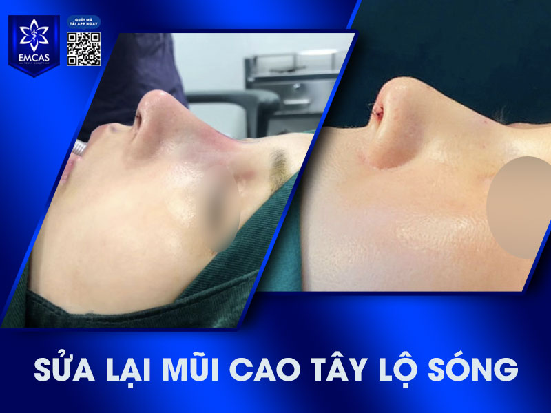
EMCAS AESTHETIC HOSPITAL
Address: 14/27 Hoàng Dư Khương Street, Hòa Hưng Ward, Ho Chi Minh City, Vietnam
Hotline: 0903 082 836 – 0979 223 636
Website: https://www.emcas.vn/en